
Fig. 3.
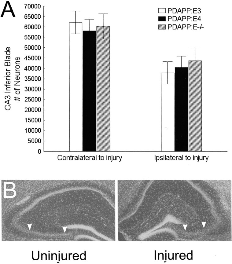
A, No group differences were found for neuronal loss within the inferior blade of CA3. B, Photomicrograph shows a DAPI-stained brain section revealing atrophy of the CA3 region (arrowheads delineate the borders of the CA3 inferior blade) 3 months after TBI.