Fig. 1.
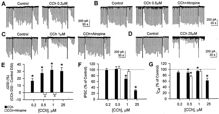
CCh enhances DSI and depresses eIPSC amplitudes. eIPSCs were evoked every 4 sec, and cells were depolarized to 0 mV from the holding potential of −70 mV every 88 sec. A–D, Representative traces from four different cells. Two DSI trials per condition are shown. CCh enhanced DSI at 0.2–25 μm and reduced eIPSC amplitude as well at ≥1 μm. The antagonistic effect of atropine (1 μm) was tested with 0.5 and 1 μm CCh. E, Changes in DSI (ΔDSI) were calculated by subtracting control DSI from DSI with CCh (filled bars). DSI with CCh was greater than control DSI at 0.2, 0.5, 1, and 25 μmCCh (n = 7, 7, 11, and 5, respectively) (*p < 0.01; paired t test). At 0.5 μm (n = 5) and 1 μm(n = 5) CCh, 1 μm atropine reversed the effect of CCh (open bars; paired ttest after repeated measures ANOVA; p > 0.1).F, eIPSC amplitude was not changed by 0.2 and 0.5 μm CCh (paired t test;p > 0.1) but reduced by 1 and 25 μm(*p < 0.001; paired t test;filled bars). Atropine (1 μm) recovered the eIPSC amplitude reduced by 1 μm CCh (open bars; paired t test after repeated measures ANOVA; p > 0.1). G, Peak Ca2+ current activated by 0 mV pulse. In the presence of 0.2–1 μm CCh, the mean Ca2+ currents were 88–94% of control (filled bars). In the presence of 0.5 μm CCh plus atropine, the Ca2+ current showed more rundown (75 ± 4% of control; open bars). CCh (25 μm) reduced the eIPSC amplitude to 61 ± 8% of control. *p < 0.05; pairedt test between control and CCh.

