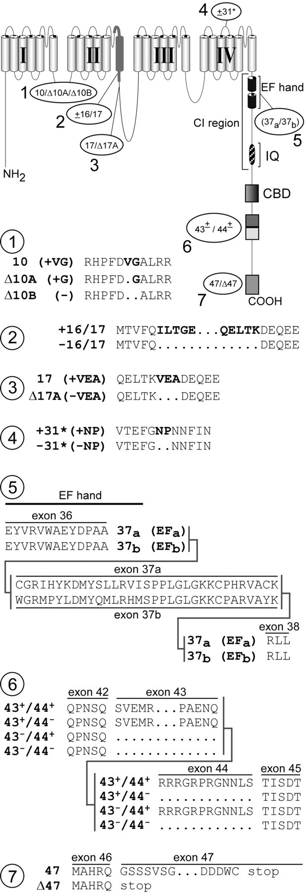
Fig. 2.

Seven loci of α12.1 splice variation detected by transcript scanning. The postulated schematic diagram (top) shows a more detailed secondary structure of α12.1, along with loci of splice variation (1–7), labeled according to transcript variant names. CI, Ca2+ inactivation region, containing structures believed important for CDI such as the EF-hand and IQ domain. Detailed changes in amino acid composition resulting from splice variation at each of seven loci are shown below. Atlocus 3, deletion of exons 16 and 17 would remove half the P-loop and the entire IIS6 segment, ostensibly producing a nonfunctional channel.
