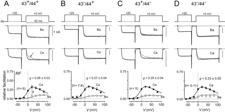
Fig. 4.
Splicing at exons 43 and 44 does not affect CDF properties. A–D, Top, Prepulse voltage protocol used to reveal facilitation, with fixed test pulse depolarization to 0–5 mV and 30 msec prepulse depolarization.Middle, Exemplar Ba2+ and Ca2+ current traces corresponding to specific voltage pulses diagrammed at top. Thegray trace corresponds to the trial without a prepulse. The arrow in A marks slow activation phase characteristic of CDF. Bottom, Population averages (from n cells) of strength of CDF (RF) plotted as a function of prepulse potential. Ca2+ data are plotted as filled symbols; Ba2+ data are plotted asopen symbols. g is a metric for the strength of pure Ca2+-dependent facilitation, and mean values and SEM are shown.

