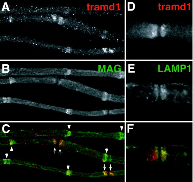
Fig. 9.
Localization of tramdorin1 in myelinating Schwann cells. Images of unfixed teased fibers from adult rat sciatic nerve, double labeled with a rabbit tramdorin1 antiserum (A,D) (TRITC) and a mouse monoclonal antibody against MAG (B) (FITC) or LAMP1 (E) (FITC) are shown. Tramdorin immunoreactivity is found at paranodes (arrows) and most incisures (arrowheads), colocalizing with MAG (A–C), and in puncta along the outer surface. The failure to detect tramdorin in all incisures is more likely the result of technical problems of antibody penetration than of heterogeneity of expression. At paranodes (D–F), tramdorin1 immunostaining does not appear to colocalize with that of LAMP1, a lysosomal marker. Both markers possess cone-like expression patterns in the paranode, in which the tramdorin expression domain is nested within the LAMP1 expression domain.

