Fig. 5.
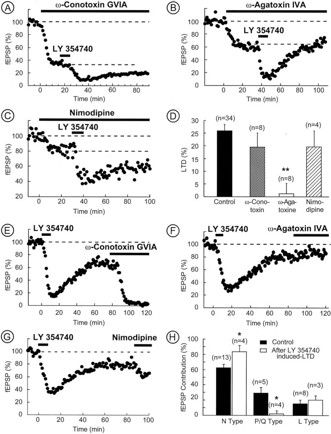
mGlu2/3-LTD is attributable to the reduction of P/Q-type Ca2+ channels to evoked synaptic transmission. A–D, The mGlu2/3-LTD requires P/Q-type Ca2+ channels. A, Typical experiment in which the slice was perfused with ω-conotoxin-GVIA (1 μm). The fraction of synaptic transmission insensitive to ω-conotoxin-GVIA displayed a normal form of mGlu2/3-LTD.B, Typical experiment in which the slice was perfused with ω-agatoxin-IVA (200 nm). The fraction of synaptic transmission insensitive to ω-agatoxin-IVA did not display mGlu2/3-LTD. C, Typical experiment in which the slice was perfused with nimodipine (1 μm). This treatment had no effect on the mGlu2/3-LTD. D, Summary of all of the experiments performed as above. The histogram of the mGlu2/3-LTD at 60 min reveals that blocking P/Q-type HVA Ca2+ channels suppressed mGlu2/3-LTD. E–H, P/Q-type Ca2+ channels do not contribute to synaptic transmission when mGlu2/3-LTD is induced. E–G, Typical experiments in which, after induction of mGlu2/3-LTD with LY354740 (200 nm), the slices were perfused with ω-conotoxin-GVIA (1 μm), ω-agatoxin-IVA (200 nm), and nimodipine (1 μm), respectively.H, Summary of all of the experiments performed as above. The histogram represents mean fEPSP inhibition (i.e., fEPSP contribution) induced by ω-conotoxin-GVIA, ω-agatoxin-IVA, and nimodipine and taken after 15–20 min of drug application. It reveals that P/Q-type Ca2+ channels do not contribute to synaptic transmission once mGlu2/3-LTD is induced. Conversely, N-type contribution is augmented. *p < 0.05; **p < 0.01.

