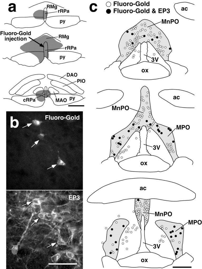
Fig. 3.

EP3 receptor-expressing POA neurons directly project to the rRPa. a, Fluoro-Gold injection centered on the caudal one-third of the rRPa (arrow). b, POA neuronal cell bodies double labeled with Fluoro-Gold fluorescence and EP3 receptor immunoreactivity (arrows). The photomicrographs were taken at the same site under different conditions of excitation.c, Distributions of POA neurons labeled with Fluoro-Gold (open circles) and with both Fluoro-Gold and EP3 receptor immunoreactivity (filled circles). All Fluoro-Gold-labeled cells distributed in the shown regions were drawn. The distribution area of EP3 receptor-immunoreactive cells is coloredgray. Scale bars: a, c, 500 μm; b, 50 μm.
