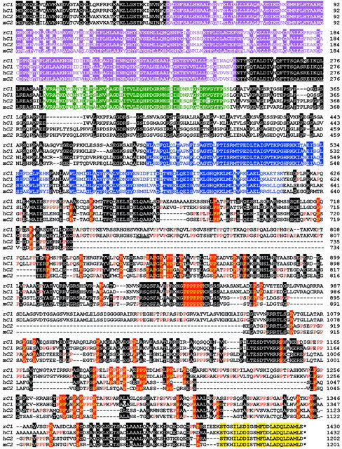
Fig. 1.

Sequence analysis of Caskins. Alignment of the primary sequences of rat and human Caskin 1 (rC1 andhC1, respectively) and human and mouse Caskin 2 (hC2 and mC2, respectively). Sequences are identified on the left and numbered on theright. Residues that are identical between Caskins 1 and 2 in at least two of the sequences shown are highlighted by a domain-specific color code: The N-terminal ankyrin repeats are shown inpurple, the SH3 domain is in green, the SAM domains are in blue, and the Caskin-specific C-terminal domain is in yellow. Outside of these defined domains, shared sequences are highlighted in blackexcept for prolines, which are shown in the C-terminal half of the proteins on a red background if conserved among different Caskins and in red typeface if specific for a given Caskin isoform.
