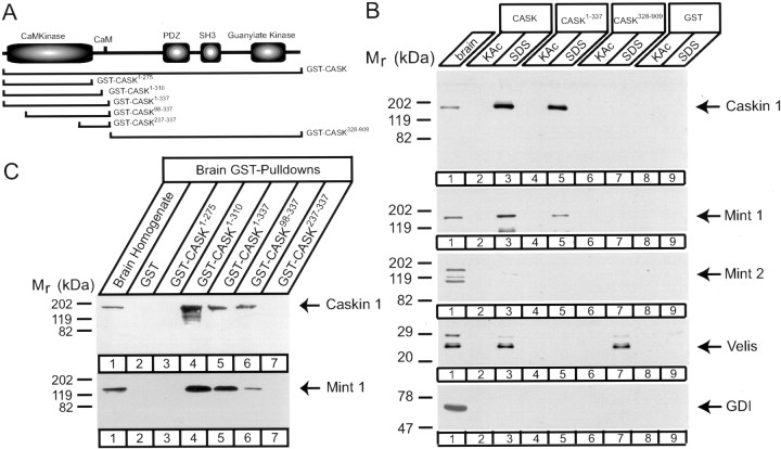
Fig. 11.
Mapping of the Caskin 1 binding site on CASK.A, Domain structure of CASK and positions of GST–CASK fusion proteins used for pulldowns. B, C, GST pulldowns of proteins in rat brain homogenates with the indicated CASK fusion proteins analyzed by immunoblotting for the proteins identified atright. B, Proteins were eluted with 0.8m K-acetate (KAc) followed by SDS sample buffer, whereas in C, proteins bound to the beads were examined. Note that in CASK, not only the CaM kinase domain but also the region homologous to the autoregulatory sequence of CaM kinase II are required for binding. Also note the separation between the common Mint 1–Caskin 1 and the Veli binding sites on CASK. Signals were visualized by ECL. Numbers at leftindicate positions of molecular weight markers.

