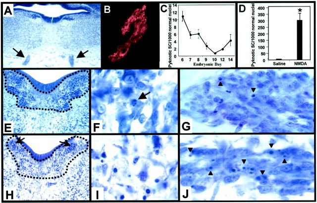
Fig. 3.
Naturally occurring and NMDA-induced Schwann cell death in the embryonic chick oculomotor nerve. The bilateral chick oculomotor nerves (arrows) exit the hindbrain and are easily accessible at E8 (A). Schwann cells of the oculomotor nerve express the avian-specific homolog of the Schwann cell specific P0 protein (B), and display a developmentally regulated period of PCD with peaks at E6 and E14 (C). NMDA treatment induces a 75-fold increase in the relative density of dying Schwann cells (D). Each point in C and D indicates the mean ± SEM for three to seven embryos. Oculomotor nuclei in E7.5 saline-treated embryos (enclosed by dotted line, E) exhibit motoneuron PCD (arrow, F), as well as Schwann cell PCD in the nerve (arrowheads, G). NMDA treatment, however, induced an excitotoxic loss of motoneurons in all oculomotor nuclei except the parasympathetic preganglionic neurons of the accessory oculomotor nucleus (arrows, H), which exhibit characteristics of a necrotic death (I), such as hyperchromatic nuclei, cytoplasmic swelling and vacuolization, and loss of Nissl substance. After NMDA administration at E7, extensive numbers of pyknotic Schwann cells (arrowheads) can be seen throughout the E7.5 oculomotor nerve (J).

