
Fig. 5.
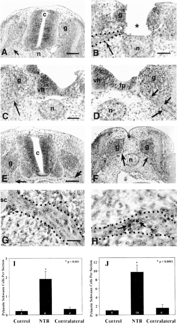
Neural tube removal at E3 in the chick embryo results in an increase in Schwann cell death in the spinal ventral roots. Compared with intact neural tubes displaying normal ventral roots (arrow, A), neural tubes surgically removed at E3, leave only the sensory ganglia (g) and ventral roots intact (arrow,B) by 2 hr after surgery. In some embryos, neural tube removal was incomplete and often resulted in one intact contralateral ventral horn with associated nerve root (arrow,C) and one ablated ipsilateral ventral horn (D), containing many pyknotic cells within its nerve root (arrows). Similar results were observed 6–8 hr (vs 1–2 hr) after neural tube removal on E3 (E–H). A control embryo with an intact neural tube (E) displays intact ventral roots (arrows), and an embryo 6–8 hr after complete neural tube removal exhibits intact sensory ganglia (g) and ventral roots (arrows,F). Little, if any, Schwann cell death is found in control embryo ventral roots (G), whereas 6–8 hr after complete neural tube removal there is a large increase in pyknotic Schwann cells (arrows,H) in the ventral roots. Quantification of pyknotic Schwann cells revealed a significant increase in the ipsilateral ventral roots as compared with both control and contralateral roots at 1–2 hr (I) and 6–8 hr (J) after neural tube removal. c, Central canal;g, sensory ganglia; n, notochord;vh, ventral horn; fp, floor plate;sc, spinal cord. In I andJ, the number in the barsrepresents sample size.