
Fig. 6.
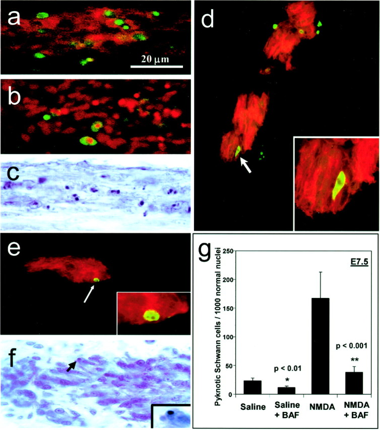
Schwann cells display characteristics of an apoptotic death in normal and NMDA-treated embryos and use a caspase-dependent pathway. Ventral root Schwann cells labeled with the 1E8 antibody (red) at E7.5, after treatment with NMDA on E7, display numerous TUNEL-positive cells (green), indicating DNA fragmentation (a). Also after NMDA treatment, dying Schwann cells are labeled with the sc45 antibody (green), which labels a downstream caspase-3 cleavage protein (b). Pyknotic Schwann cells in the ventral root, displaying darkly stained condensed nuclei are abundant in Nissl-stained tissue after NMDA treatment (c). In the oculomotor nerve, dying Schwann cells labeled with 1E8 (red) are colabeled with an antibody to activated caspase-3 (green) (d), as well as with the antibody to sc45 (green) (e). Normally dying Schwann cells in the oculomotor nerve in Nissl-stained tissue display a pyknotic morphology with darkly stained, condensed nuclear chromatin (arrow,f). Inset in fshows a pyknotic Schwann cell at higher magnification. Embryos were treated at E7 with either saline or NMDA, followed by a 40 μg dose of BAF to prevent caspase activation. Analysis at E7.5 showed significant reductions in Schwann cell death after BAF treatment in both saline-treated (p < 0.01) and NMDA-treated (p < 0.001) embryos (g). Bars represent the mean ± SEM for four to seven embryos. The scale ina is for a–f.