
Fig. 10.
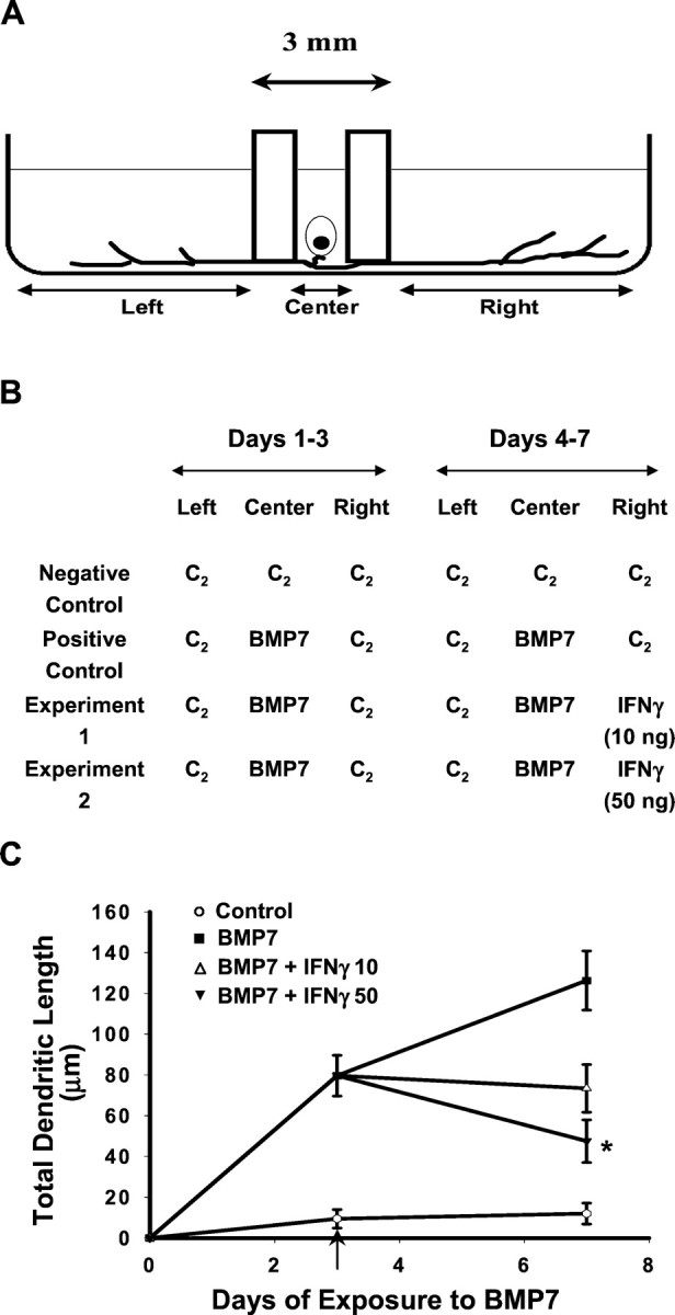
Dendritic retraction in response to retrograde signaling by IFNγ. A, Schematic representation of the three-compartment culture of sympathetic neurons. Dissociated neurons were plated in the Center compartment, and axons extended into the Left and Rightcompartments. B, Negative control cultures were maintained in control medium (C2) in the absence of BMP-7 or IFNγ; positive control and experimental cultures were exposed to BMP-7 (10 ng/ml) added to C2 in the Center compartment for 7 d. Three days after the addition of BMP-7, IFNγ (10 or 50 ng/ml) was added to the C2 in the Right compartment of experimental cultures (indicated by the arrow in C). Then 4 d later, dendritic growth was quantified in cultures immunostained with mAb to MAP-2 (n ≥ 60 cells/condition). Addition of IFNγ to the distal axons and axon terminals caused a dose-dependent reduction in total dendritic length (C). ∗p < 0.05 versus BMP-7 on day 7.