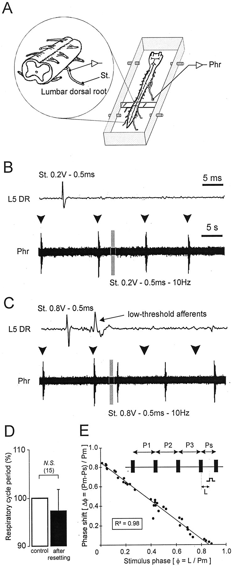
Fig. 3.

Ability of low-threshold lumbar afferents to reset spontaneous respiratory rhythmicity. A, Schematic representation of the experimental procedure. B, C, Continuous recordings of spontaneous phrenic (Phr) activity during a volley of lumbar (L5) dorsal root (DR) stimulation. Shown above each phrenic trace is a faster time base recording from the corresponding L5 DR during a single shock at the indicated stimulus intensity. The gray barindicates a train stimulation of lumbar afferents. Subthreshold electrical stimulation (0.2 V) of lumbar afferents (B) did not reset the respiratory phrenic rhythmicity, whereas respiratory resetting was obtained when low-threshold lumbar afferents were activated by ≥0.8 V (C). Arrowheads denote the expected time of occurrence of spontaneous phrenic bursts in the absence of resetting. D, Histograms showing lack of significant change in respiratory period (expressed as percentage of the mean control period) after resetting. The control value corresponds to the mean of three successive respiratory periods before the stimulated cycle (white bar), which is compared with the respiratory cycle observed after the stimulated cycle (black bar). N.S., Nonsignificant. E, Phase response plot calculated as follows (also see schematic): the reference period (Pm) was measured from three spontaneous respiratory cycles (P1, P2, and P3); the ratio of the stimulus latency (L) and Pm determined the stimulus phase (φ); the phase shift of the phrenic burst (Δφ) expressed as the difference between Pm and the stimulated period (Ps) and divided again by Pm, was plotted on the ordinate. The solid line indicates linear regression.R2, Coefficient of determination. Standardized data were collected from three different preparations.
