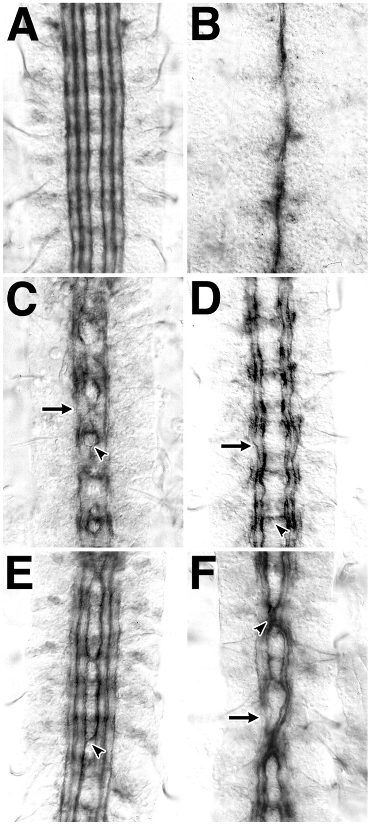
Fig. 1.

slit interacts genetically withrobo and dock. A bilateral set of three distinct axon fascicles are labeled with antibody to Fasciclin II in stage 17 embryos of wild-type Drosophila(A). All axons fuse at the midline in embryos mutant for slit (B), whereas medial axons recross the midline in embryos mutant for a Slit receptor gene, robo (C, arrowhead). Lateral axons do not make midline guidance errors, but occasional gaps in Fasciclin II labeling are seen (C,arrow). Midline guidance errors are very rare in embryos mutant for dock (D,arrowhead); however, gaps in Fasciclin II labeling in the lateral axon fascicles are common (D,arrow). Midline guidance errors in the most medial axon tract are common in embryos that have a single wild-type and a single mutant copy of both the slit and robogenes (E, arrowhead). Embryos similarly heterozygous for both slit and dock have more frequent and profound midline guidance errors, involving all labeled axon fascicles (F). In this and subsequent figures,arrowheads indicate midline guidance errors, andarrows indicate interruptions in the longitudinal tracts.
