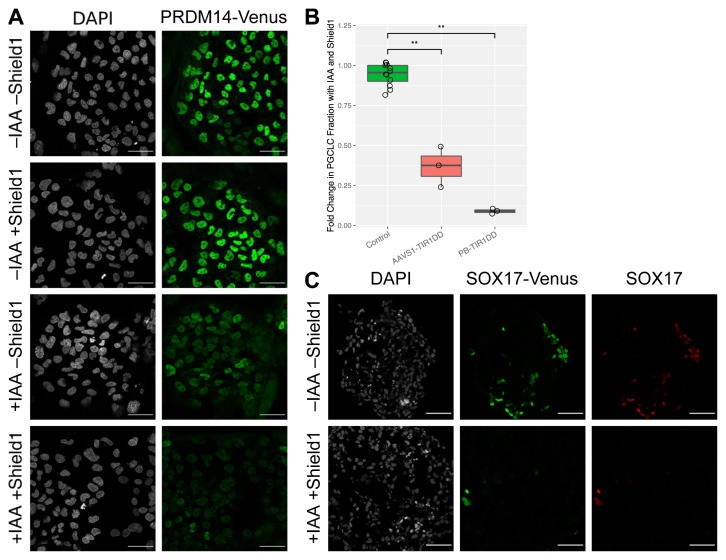
Figure 5. AID depletion of PRDM14 and SOX17 using inducible TIR1.
( A) PRDM14-AID-Venus/TIR1-DD human embryonic stem cells (hESCs) grown on a layer of mouse embryonic fibroblasts were treated with indole-3-acetic acid (IAA) and/or Shield1. After one hour of treatment, cells were fixed and stained for IF using anti-GFP. Scale bar is 50 µm. ( B) SOX17 depletion abrogates hPGCLC induction. SOX17-AID-Venus hESCs, expressing TIR1-DD either from AAVS1 or from random PiggyBac integration, were induced to form hPGCLCs with and without Shield1 and IAA. AP+/NANOS3+hPGCLC fraction was measured by flow cytometry after four days. Fractions were normalized with respect to untreated samples of the same clones. Results from cells without TIR1 are plotted for comparison. Significance values are by Wilcoxon test (*p < 0.05, **p < 0.01, ***p < 0.001). Each point represents a biological replicate. ( C) SOX17 depletion by AID with TIR1-DD. SOX17-AID-Venus cells with PB-TIR1-DD were induced to form human primordial germ cell-like cells (hPGCLCs) and treated with IAA and Shield1 from the start of the induction. After four days, EBs were fixed, sectioned, and stained using antibodies against GFP and SOX17. The untreated sample contains many SOX17 positive hPGCLCs, whereas only a few are visible in the treated sample. Scale bar is 50 µm.

