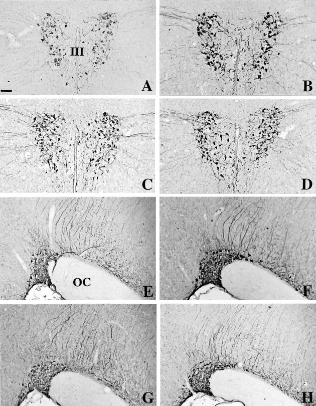
Fig. 1.

Immunohistochemical detection of arginine-vasopressin in the PVN (A–D) and in the SON (E–H) of C3H mice (A, E), Tg8 mice (B, F), α-MPT-treated Tg8 mice (C, G), and pCPA-treated Tg8 mice (D, H). The mutation is associated with an increase in AVP immunoreactivity in the PVN (B vs A) and in the SON (F vs E). No difference was observed between Tg8 and saline-control Tg8 mice. The treatment by α-MPT in Tg8 mice is correlated with a decline in AVP immunoreactivity in the PVN (C) as well as in the SON (G) compared with saline-control Tg8 mice (the same as B, F). In pCPA-treated Tg8 mice the intensity of labeling is also decreased in the PVN (D) and in the SON (H) compared with saline-control Tg8 mice.III, Third ventricle; OC, optic chiasma. Scale bar, 50 μm.
