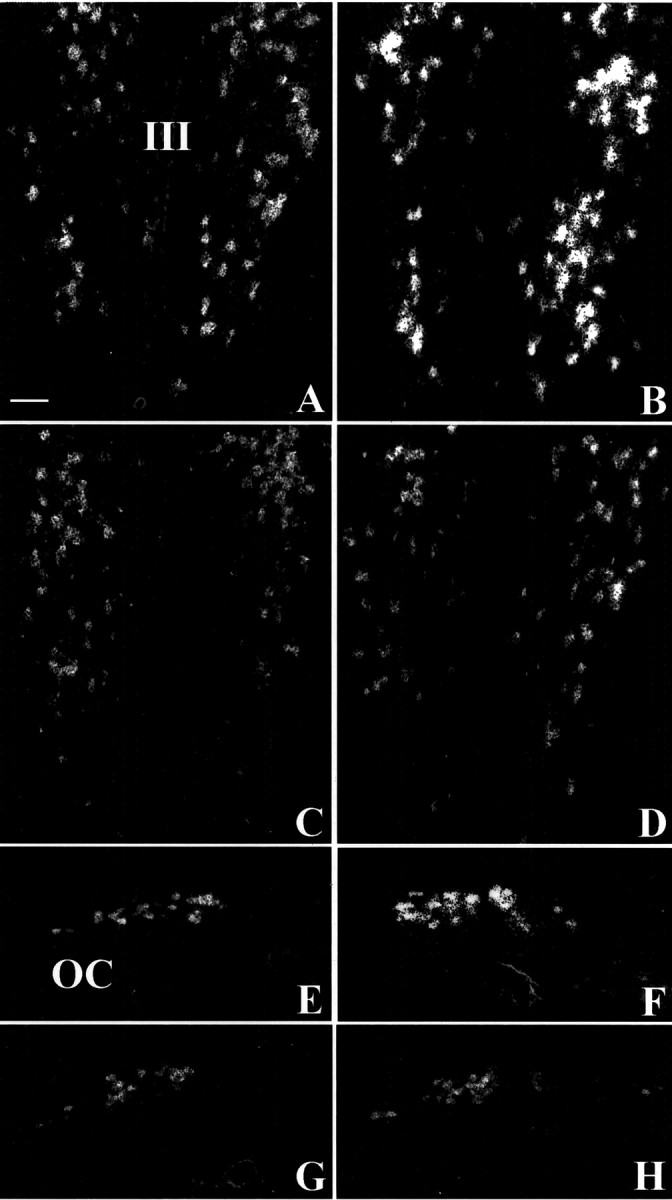
Fig. 4.

Dark-field microphotographs representing thein situ hybridization signal of OT mRNA on emulsion-coated sections in C3H mice (A, E), Tg8 mice (B, F), α-MPT-treated Tg8 mice (C, G), and pCPA-treated Tg8 mice (D, H). The hybridization signal is enhanced in Tg8 mice both in the PVN (B) and the SON (F) compared with C3H mice (A, E). In α-MPT-treated Tg8 mice the hybridization signal is reduced in the PVN (C) as well as in the SON (G) compared with saline-control Tg8 mice (the same as B, F). In pCPA-treated Tg8 mice the density of silver grains is also decreased in the PVN (D) and in the SON (G) compared with saline-control Tg8 mice. III, Third ventricle; OC, optic chiasma. Scale bar, 50 μm.
