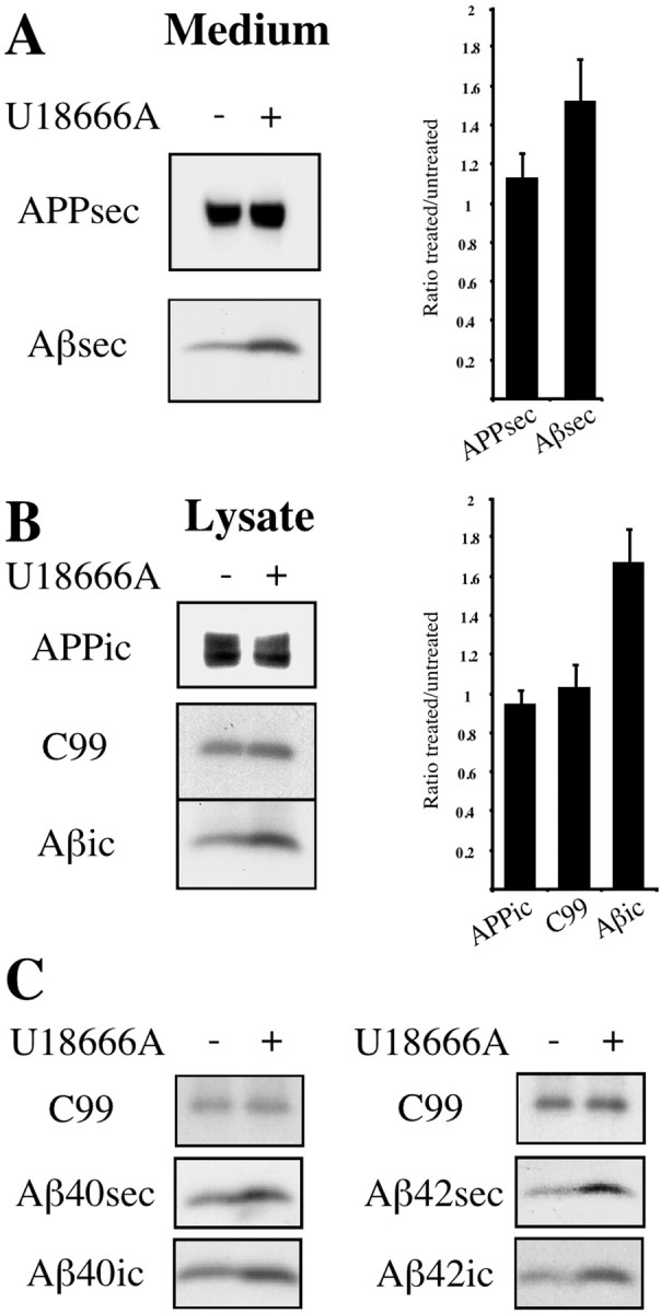
Fig. 2.

U18666A increases intracellular and secretory Aβ levels in SP-C99-transfected SH-SY5Y cells. Human SH-SY5Y neuroblastoma cells stably transfected with APP C-terminal fragment SP-C99 were incubated for 24 hr with 50 μg/ml LDL in the presence (+) or absence (−) of 3 μg/ml U18666A. Conditioned medium (A) and cell lysates (B) were immunoprecipitated with antibody W02, followed by Western blot detection with W02. Graphs show quantification of intracellular (ic) and secreted (sec) endogenous APP, C99, and overall Aβ levels (n = 5 experiments). Depicted are ratios of signal intensities from U18666A-treated versus untreated cells. Error bars indicate 1 SD. C, Medium and lysates of SH-SY5Y cells were immunoprecipitated with antibodies G2-10 and G2-11 specific for Aβ40 and Aβ42, respectively, and were detected with W02. Western blots with W02 show effects of U18666A treatment on secretory and intracellular Aβ species compared with C99.
