
Fig. 4.
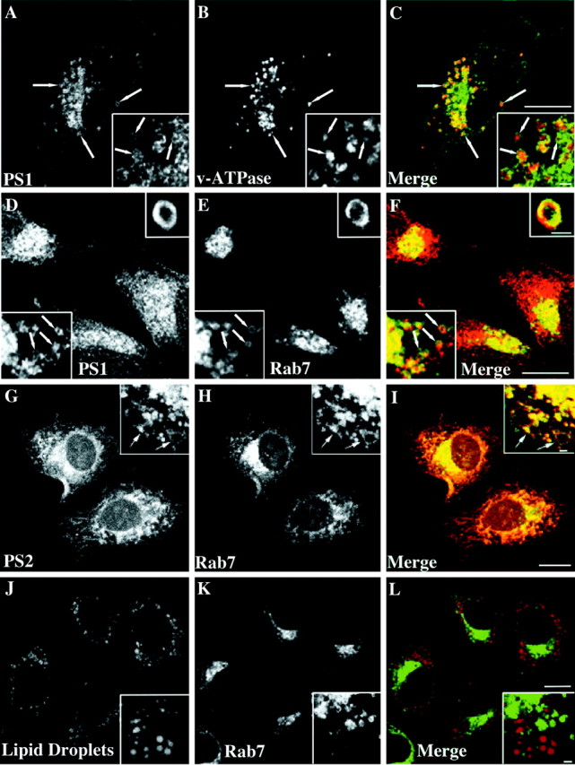
PS-containing compartments are positive for Rab7 and surround late endosomes in a ring-like manner. SH-SY5Y (A–C) or A431 cells (D–L) were incubated for 24 hr with 50 μg/ml LDL and 3 μg/ml U18666A. After fixation in paraformaldehyde the SH-SY5Y cells were stained against PS1 (A, C; green) and v-ATPase (B, C; red). Fixed A431 cells stably overexpressing Rab7-GFP (E, H, K;green) were labeled with a monoclonal PS1 antibody (D, F; red), polyclonal PS2 antibody (G, I; red), or Oil red O to visualize neutral lipids (J, L; red). C, F, I, L, Merged images. Scale bars, 8 μm. Arrowsindicate selected vesicles positive for both markers.Insets show single sections from confocal stacks of random vesicular structures in cell bodies. Scale bars ininsets, 1 μm.