Fig. 1.

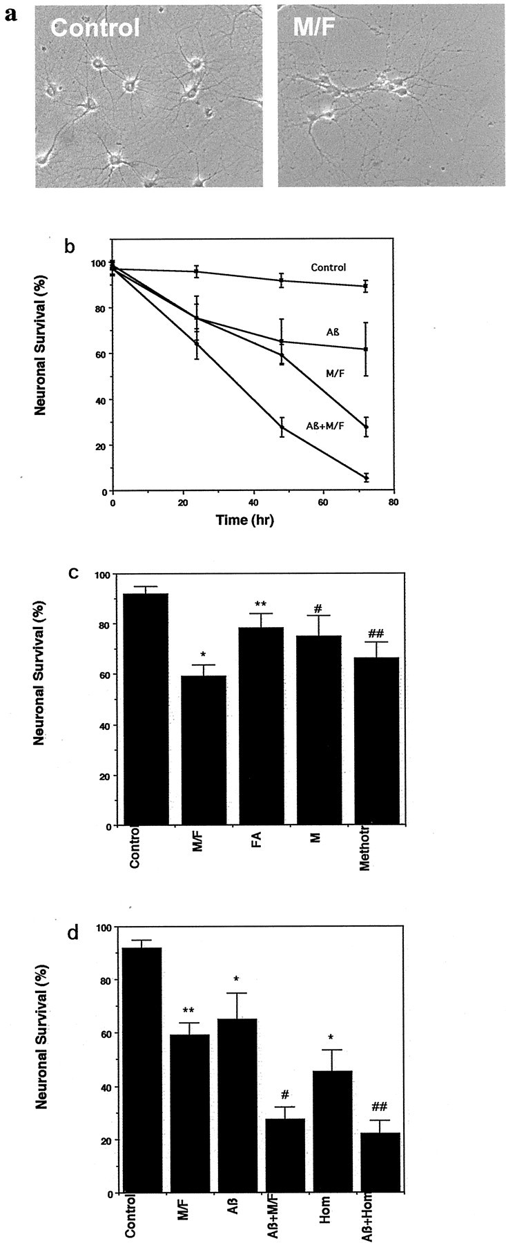
Methyl donor deficiency induces death of hippocampal neurons and potentiates Aβ toxicity. a, Phase-contrast micrographs showing hippocampal neurons in a control culture and a culture that had been maintained for 48 hr in medium lacking methionine and folic acid. b, Cultures were exposed to control medium, medium lacking l-methionine and folic acid (M/F), medium containing 5 μm Aβ1-42 (Aβ), or medium containing a combination of M/F plus Aβ; neuronal survival was quantified at the indicated time points. Neuronal survival is expressed as a percentage of the initial number of neurons present before experimental treatment (see Materials and Methods). Values are the mean and SD of determinations made in six cultures. c, Cultures were incubated for 48 hr in control medium, medium lacking methionine and folic acid (M/F), medium lacking folate (FA), medium lacking methionine but containing folic acid (M), or medium containing 20 μm methotrexate (Methotr). Neuron survival was quantified (mean and SD; n = 6). *p < 0.001 compared with control; **p < 0.01 compared with M/F;#p < 0.05 and##p < 0.01 compared with control (ANOVA with Scheffe's post hoc tests).d, Cultures were exposed for 48 hr to saline (Control), M/F-deficient medium, 5 μm Aβ1-42 (Aβ), a combination of M/F-deficient medium plus 5 μm Aβ1-42 (Aβ+M/F), 250 μm homocysteine (Hom), or a combination of homocysteine plus Aβ (Aβ+Hom). Neuron survival was quantified (mean and SD;n = 6). *p < 0.01, **p < 0.001 compared with control;#p < 0.01 compared with M/F deficiency and with Aβ; ##p < 0.01 compared with homocysteine and with Aβ (ANOVA with Scheffe's post hoc tests).
