
Fig. 2.
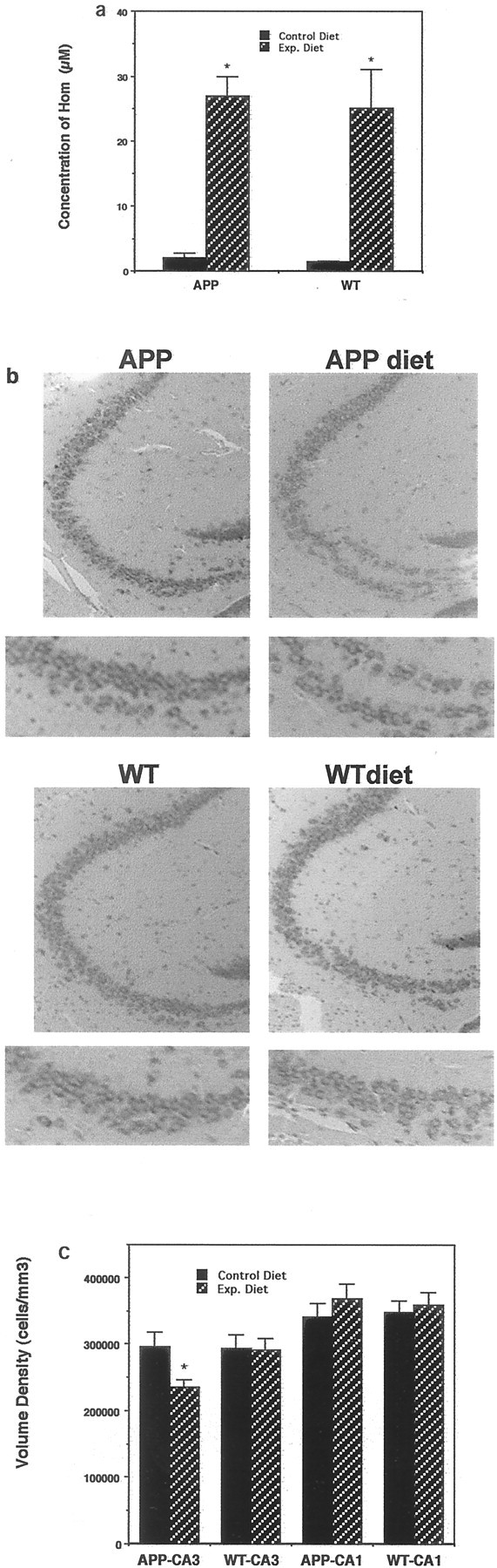
A methyl donor-deficient diet induces hyperhomocysteinemia and promotes neuronal degeneration in APP mutant mice. a, Levels of homocysteine in serum samples from wild-type (WT) and APP mutant mice that had been maintained for 3 months on the normal control diet or the experimental folic acid-deficient diet were quantified. Values are the mean and SD (n = 8). *p < 0.0001 compared with corresponding control value (ANOVA with Scheffe's post hoc tests). b, Micrographs showing cresyl violet-stained sections of hippocampus (region CA3) from wild-type and APP mutant mice maintained for 3 months on either the control diet or the folic acid-deficient diet (APP diet; WT diet). The micrographs at thebottom show high magnification of the lower limb of CA3.c, Numerical densities of neurons in regions CA3 and CA1 of hippocampus were quantified in the brains of wild-type and APP mutant mice maintained for 3 months on either control or folic acid-deficient diets. Values are the mean and SD (n= 8). *p < 0.01 compared with APP mutant or wild-type mice maintained on the control diet and compared with wild-type mice on the experimental diet (ANOVA with Scheffe'spost hoc tests).