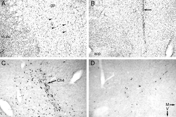
Fig. 1.

A, B, Photomicrographs of sections of the basal forebrain stained with cresyl violet showing the effects of infusion of 192 IgG-saporin (SAP HIGH) (B) compared with a control subject that received infusions of vehicle at the same coordinates (A). The magnocellular, hyperchromatic neurons of the nucleus basalis neurons that can be seen in A(arrowheads) are markedly reduced in number in the SAP-infused brain (B), despite otherwise excellent preservation of neurons in the globus pallidus (gp) and ventrolateral caudate-putamen (vlcpu). The arrow in Bindicates the glial-rich site of the cannula tract through which 192 IgG-saporin infusions were made. C, D, Sections adjacent to A and B, respectively, stained using antibodies to choline acetyltransferase to reveal the magnocellular neurons of the Ch4 cell group (C, arrow). The marked reduction in the number of cholinergic neurons can be seen in the 192 IgG-saporin-infused brain (SAP HIGH) (D).acp, Posterior limb of the anterior commissure;M, medial; V, ventral.
