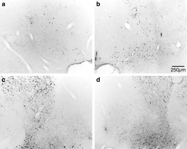
Fig. 2.
Representative photomicrographs showing ChAT-IR (a, b) and PARV-IR (c, d) neurons in the basal forebrain of SHAM (right-hand panels) and SAP HIGH (left-hand panels) lesioned rats. It can be seen that the magnocellular ChAT-IR neurons of the Ch4 cell group (nucleus basalis magnocellularis) are greatly reduced in number after intrabasalis infusions of 192 IgG-saporin. By contrast, parvalbumin-containing neurons are unaffected by immunotoxin infusions in this region where cholinergic neurons are lost (comparea, c).

