
Fig. 4.
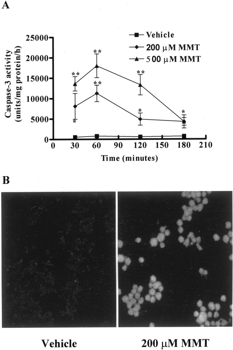
MMT treatment increases caspase-3 activity.A, Caspase-3 enzymatic activity. B, In situ caspase-3 activity. A, Subconfluent cultures of undifferentiated PC12 cells were harvested at 30 min, 1, 2, and 3 hr after MMT treatment. Caspase-3 activity was assayed using specific fluorogenic substrate, Ac-DEVD-AMC (50 μm), as described in Materials and Methods. The data represent mean ± SEM of nine individual measurements from three separate experiments. Asterisks (**p < 0.01; *p < 0.05) indicate significant differences compared with temporally matched vehicle (DMSO)-treated cells. B, PC12 cells were grown on laminin-coated slides for 2–3 d and then exposed to 0.5% DMS0 (vehicle) and 200 μm MMT for 1 hr in the dark. After exposure, cells were treated with 10 μm FITC-VAD-FMK (Promega caspACE, in situ marker for caspase-3 activity) and processed as described in Materials and Methods. Confocal images were obtained using a Leica TCS-NT microscope.