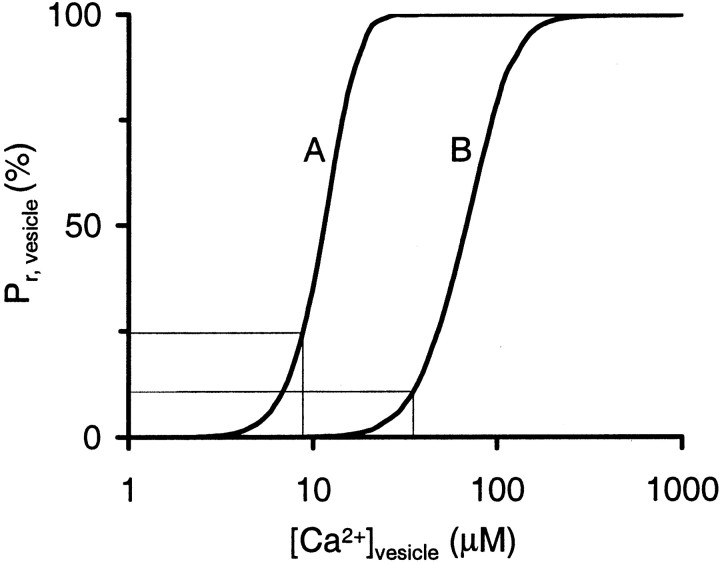
Fig. 1.
Intrinsic Ca2+ sensitivity of transmitter release. Shown is Pr for a single vesicle when exposed to a [Ca2+] transient with a time course equal to that of whole-cellICa (full width at half-maximum, 383 μsec) and with a peak amplitude of [Ca2+]vesicle. The thin lines indicate release probability during APs according to Release Model A (Pr, vesicle = 25% at [Ca2+]vesicle = 8.8 μm; Bollmann et al., 2000) or Release Model B (Pr, vesicle = 10% at [Ca2+]vesicle = 35 μm; Schneggenburger and Neher, 2000).

