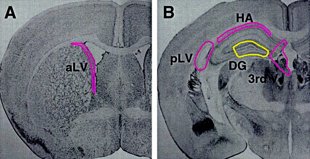
Fig. 1.
Dissection and rate of sphere formation of adult mouse neurogenic regions. A, B, Atlas images of adult mouse brain sections (Franklin and Paxinos, 1997) adapted to show the microdissection of viable 500 μm vibratome sections used to isolate tissue from neurogenic regions.A, Coronal section through the anterior lateral ventricle (aLV) with the dissected region highlighted. Note that this dissection includes both subependymal and ependymal tissue, but for the purposes of this study ependymal sphere formation was ignored. B, Coronal section through the hippocampus. Note that the dentate gyrus (DG) dissection excludes all regions containing subependymal tissue; these regions were dissected and cultured separately. 3rd V, Third ventricle; pLV, posterior lateral ventricle;HA, hippocampal arch. This dissection scheme was used for both rats and mice.

