Figure 1.
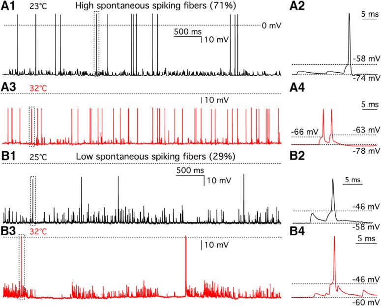
Heterogeneity in the temperature dependence of afferent fiber spikes. A1, Whole-cell current-clamp recordings of afferent fiber with zero current injection at 23°C. AP spikes and EPSPs can be clearly distinguished. Dashed line indicates 0 mV. A2, The spike shown in a dashed line box in A1 was expanded in time scale. Resting membrane potential (Vrest) was −74 mV, and AP threshold was −58 mV. A3, Spontaneous spikes from the same afferent fiber shown in A1 were recorded at 32°C. A4, Spikes shown in a dashed line box in A3 are expanded in time scale. Vrest was −78 mV. Thresholds of the first and the second AP were −66 and −63 mV, respectively. Five of seven afferent fibers (71%) fired more spikes at high temperature. B1, Spontaneous spikes were recorded from another afferent fiber at 25°C. Dashed line indicates 0 mV. B2, The spike shown in a dashed line box in B1 was expanded in time scale. Vrest was −58 mV, and AP threshold was −46 mV. B3, Spontaneous spikes from the same fiber shown in B1 were recorded at 32°C. B4, The spike shown in a dashed line box in B3 was expanded in time scale. Vrest was −60 mV, and AP threshold was −46 mV. Two of seven afferent fibers (29%) fired less spikes at high temperature.

