Figure.

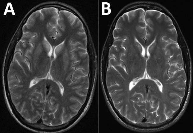
Magnetic resonance imaging (MRI) of the brain of a patient with encephalitis caused by Powassan virus, Massachusetts, USA, 2017. A) Initial brain MRI showing high T2 signal abnormality in the bilateral caudate and putamen. B) Noticeable improvement on repeat brain MRI 2 weeks later.
