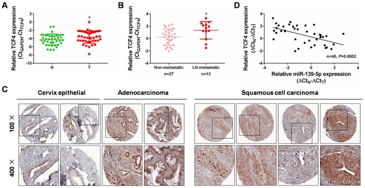
Figure 6.
Increased expression of TCF4 and its inverse correlation with miR-139-5p expression in CC. (A) qRT-PCR analysis was performed to detect TCF4 expression in 40 pairs of cervical cancer tissues and matched non-cancerous tissues. N: normal, T: tumor. (B) qRT-PCR analysis was performed to detect TCF4 expression in lymph node metastatic CC tissues (n=13) compared with non-metastatic CC tissues (n=27). LN: lymph node. (C) Immunohistochemistry analyses from the online tool human protein atlas (https://www.proteinatlas.org/) validated TCF4 protein upregulation in CC tissues. Up panel: 100×, down panel: 200×. (D) Spearman’s correlation analysis was performed to explore the negative correlation between miR-139-5p and TCF4 mRNA levels in paired CC tissues (n=40, P=0.0002). *P<0.05.

