FIG 12.
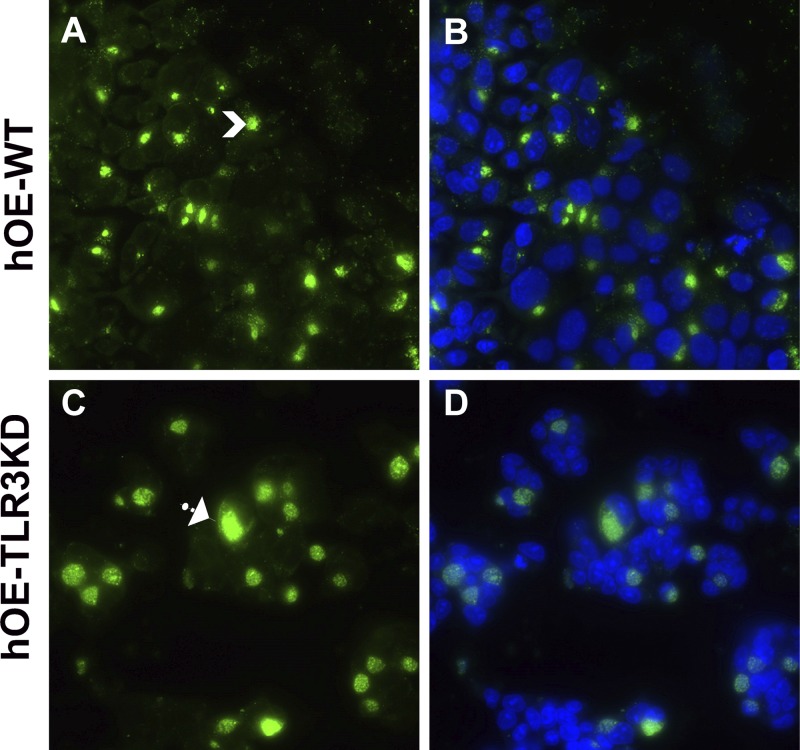
TLR3 deficiency in murine OE cells leads to large and aberrantly shaped chlamydial inclusions. hOE-WT cells (A and B) and hOE-TLR3KD cells (C and D) were either mock treated or infected with C. trachomatis serovar D at an MOI of 10 IFU/cell for 36 h. Chlamydial inclusions were stained using anti-chlamydial LPS monoclonal antibody and detected via an Alexa Fluor 488-conjugated secondary antibody. Nuclei were visualized via DAPI staining (B and D). Data shown are representative. Arrows show smaller versus larger inclusions. Magnification, ×60.

