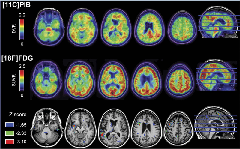
Figure 3.

Top row depicts the patient’s PiB-PET distribution volume ratio (DVR) image, which was initially read as equivocal for amyloid binding at the time of Ms. X’s research visit. However, this is now visually read as an early positive scan. Cortical binding can be observed mainly in the precuneus and anterior cingulate cortex. Middle row shows the patient’s FDG-PET standardized uptake value ratio (SUVR). Bottom row illustrates the comparison of the patient’s FDG-SUVR to a control group of clinically normal age-matched women (n = 13, age at FDG: 66.6 ± 2.7). Colored areas correspond to voxels where the patients’ SUVR value was significantly lower than the controls (corresponding to uncorrected one-tailed p < 0.05, 0.01, and 0.001), confirming a left-asymmetric pattern hypometabolism in left operculum and superior temporal gyrus regions
