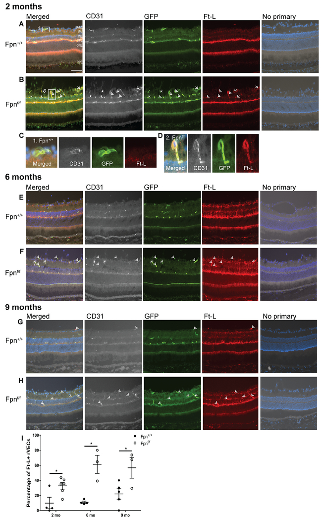
Figure 2. Fpn-deletion in the r&bVECs leads to Ft-L accumulation in GFP+ blood vessels in the retina.
At the 2 mo time point, there was increased Ft-L labeling within the GFP+ rVECs (rVECs were identified by co-labeling with CD31) of Fpnf/f experimental mice (B) compared to age-matched controls (A). (C): A magnified image of a Ft-L−, GFP+ rVEC from the 2 mo Fpn+/+ control retina. (D): A magnified image of a Ft-L+, GFP+ rVEC from the 2 mo Fpnf/f experimental retina. At the 6 mo time point, there was increased Ft-L labeling within the GFP+ rVECs of experimental mice (F) compared to age-matched controls (E). At the 9 mo time point, there was increased Ft-L labeling within the GFP+ rVECs of experimental mice (H) versus controls (G). Scale bar, 50 μm. Quantification of percentage of Ft-L positive, GFP+ blood vessels in retinas at three time points (I). White arrows point to Ft-L+, CD31+, GFP+ rVECs. * p<0.05, *** p<.001. Abbreviations: GCL, ganglion cell layer; IPL, inner plexiform layer; INL, inner nuclear layer; OPL, outer plexiform layer; ONL, outer nuclear layer; RPE, retinal pigment epithelium.

