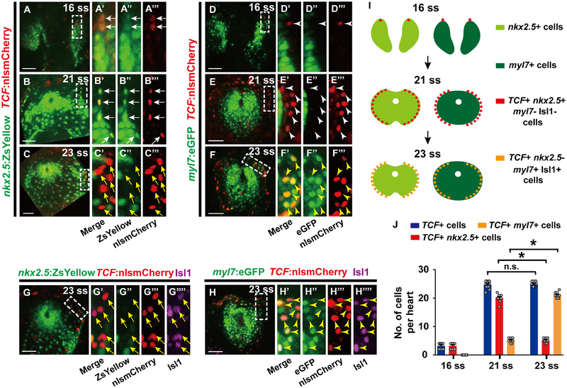
Figure 2. Nkx2.5 expression is silenced while canonical Wnt signaling is activated in nkx2.5+ progenitors during pacemaker cardiomyocyte differentiation.
(A-F’’’) Confocal images of (A-C’’’) Tg(nkx2.5:ZsYellow; TCF:nlsmCherry) (n = 8 embryos per stage) or (D-F’’’) Tg(myl7:eGFP; TCF:nlsmCherry) embryos (n = 8, 8, 10 embryos for each respective stage) at (A-A’’’, D-D’’’) 16 ss, (B-B’’’, E-E’’’) 21 ss, or (C-C’’’, F-F’’’) 23 ss reveal that canonical Wnt signaling is activated in outlying Nkx2.5+ mesodermal cells which are decreasing nkx2.5:ZsYellow and increasing myl7:eGFP expression. (G-H’’’’) Anti-Isl1 immunostaining of (G-G’’’’) Tg(nkx2.5:ZsYellow; TCF:nlsmCherry) (n = 9 embryos) and (H-H’’’’) Tg(myl7:eGFP; TCF:nlsmCherry) (n = 10 embryos) embryos at 23 ss shows that canonical Wnt signaling (TCF:nlsmCherry+) is activated in nkx2.5:ZsYellow- Isl1+ cells and myl7:eGFP+ Isl1+ cardiomyocytes. (A’-A’’’, B’-B’’’, C’-C’’’, D’-D’’’, E’-E’’’, F’-F’’’, G’-G’’’’, H’-H’’’’) Insets are magnifications of boxed areas in A, B, C, D, E, F, G, H, respectively. Images A’’-A’’’, B’’-B’’’, C’’-C’’’, D’’-D’’’, E’’-E’’’, F’’-F’’’, G’’-G’’’’, H’’-H’’’’ are single channels from A’, B’, C’, D’, E’, F’, G’, H’ merged images, respectively. (I) Schematics based on A-H’’’’ illustrate the dynamic changes in expression of nkx2.5:ZsYellow, myl7:eGFP, Isl1, and canonical Wnt-activation (TCF:nlsmCherry+) during pacemaker cardiomyocyte differentiation. (J) Further supporting these changes, quantification of TCF:nlsmCherry+ cells, TCF:nlsmCherry+ nkx2.5:ZsYellow+ cells and TCF:nlsmCherry+ myl7:eGFP+ cardiomyocytes (per heart) at 16 ss, 21 ss and 23 ss reveals that TCF:nlsmCherry+ nkx2.5:ZsYellow+ cells are significantly reduced while TCF:nlsmCherry+ myl7:eGFP+ cardiomyocytes are greatly increased from 21–23 ss. However, the total number of TCF:nlsmCherry+ cells is not changed. White arrows point to TCF:nlsmCherry+ nkx2.5:ZsYellow+ cells. Yellow arrows point to TCF:nlsmCherry+ nkx2.5:ZsYellow- cells. White arrowheads point to TCF:nlsmCherry+ myl7:eGFP- cells. Yellow arrowheads point to TCF:nlsmCherry+ myl7:eGFP+ cardiomyocytes. Green – (A-A’’, B-B’’, C-C’’, G-G’’) nkx2.5:ZsYellow, (D-D’’, E-E’’, F-F’’, H-H’’) myl7:eGFP; Red – (A, A’, A’’’, B, B’, B’’’, C, C’, C’’’, D, D’, D’’’, E, E’, E’’’, F, F’, F’’’, G, G’, G’’’, H, H’, H’’’) TCF:nlsmCherry; Magenta – (G, G’, G’’’’, H, H’, H’’’’) anti-Isl1 immunostaining. Scale bar, 50 μm. Mean ± s.e.m. *P< 0.05 by Student’s t-test. n.s. - not significant. See also Figure S2.

