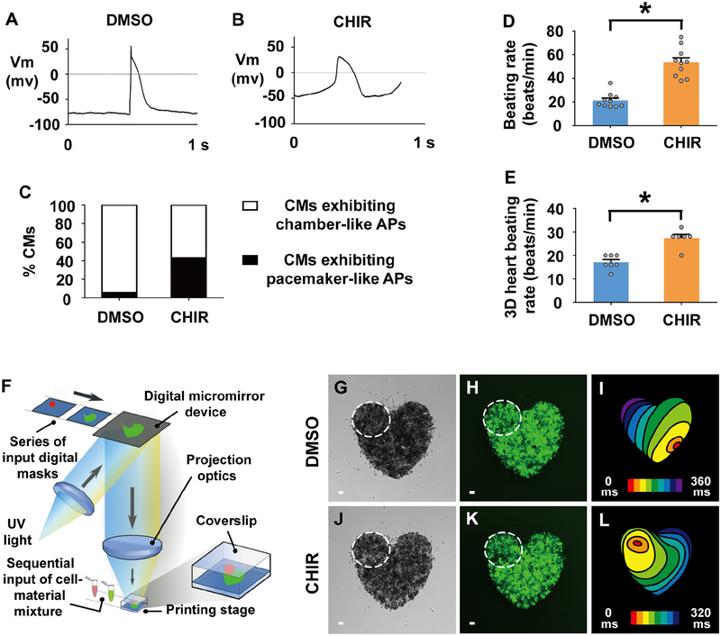
Figure 7. Activating canonical Wnt signaling in human pluripotent stem cell (hPSC)-cardiac progenitors creates functional pacemaker-like cardiomyocytes that can pace hPSC-cardiomyocytes in vitro.
(A, B) Electrophysiology recordings reveal action potentials of individual cardiomyocytes sorted from D15 cultures which were treated with (A) DMSO (n = 18 cardiomyocytes) or (B) CHIR (n = 14 cardiomyocytes) from D5–7. (C) Electrophysiology studies on these TNNT2:GFP+ hPSC-cardiomyocytes (CMs) reveals that a greater percentage of CHIR-treated hPSC-cardiomyocytes (n = 14 cardiomyocytes) exhibit pacemaker-like action potentials (APs) than DMSO-treated hPSC-cardiomyocytes (n = 18 cardiomyocytes). (D) Bar graph shows the average beating rate for cardiomyocytes sorted from D15 cultures which were exposed to either DMSO (n = 10 cardiomyocytes) or CHIR (n = 10 cardiomyocytes) treatment from D5–7. (E-L) hPSC-derived pacemaker-like cardiomyocytes can pace hPSC-cardiomyocyte tissue in vitro. (F) Diagram illustrates 3D bioprinting approach. (G-L) To create multi-cellular in vitro mini-hearts, D15 hPSC-cardiomyocytes treated with (G, H) DMSO or (J, K) CHIR from D5–7 are printed in the upper left region of mini-hearts (dashed lines encircle area), and D15 hPSC-cardiomyocytes are printed outside this encircled region. (G, J) Bright-field and (H, K) GFP microscopy images show printed mini-hearts. (I, L) Optical mapping of the calcium activation in these mini-hearts reveals that cardiac conduction is stably initiated from the site of printed (L) CHIR-treated pacemaker-like cardiomyocytes (n = 6 mini-hearts) but not (I) DMSO-treated control cardiomyocytes (n = 7 mini-hearts). Red/orange – cardiac conduction initiation site; Isochronal lines – 40 milliseconds (ms) intervals. (E) Bar graph shows that the average beating rate is significantly faster in mini-hearts printed with CHIR-treated pacemaker-like cardiomyocytes (n = 6 mini-hearts) compared to those printed with DMSO-treated control cardiomyocytes (n = 7 mini-hearts). Scale bar, 50 μm. Mean ± s.e.m. *P< 0.05 by Student’s t-test. See also Videos S1–S4.

