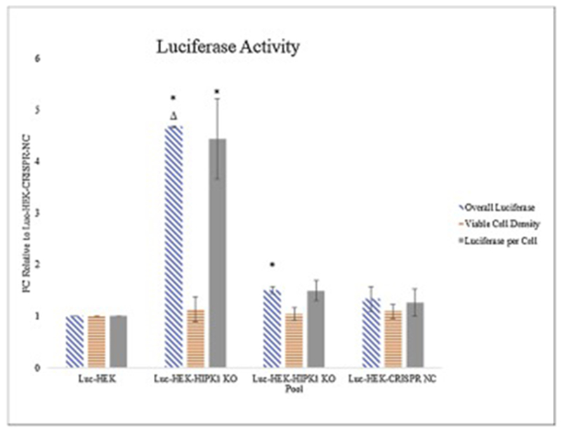
Figure 3:

Effect of knock-out of HIPK1 in luciferase-expressing HEK cells
Fold change (FC) of the overall luciferase (blue), cell viability (orange), and luciferase per cell (grey) of Luc-HEK, Luc-HEK-HIPK1 KO, Luc-HEK-HIPK1 KO pool, and Luc-HEK-CRISPR-NC cells relative to Luc-HEK demonstrates improved luciferase activity with knockout of HIPK1. Error bars represent Standard Error of the Mean (SEM) from triplicate measurements. * indicates P ≤ 0.05 relative to Luc-HEK and Δ indicates P ≤ 0.05 relative to Luc-HEK-CRISPR-NC calculated using two-sample unpaired t-Test assuming unequal variances.
