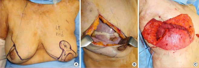
Fig. 2. Preoperative plan and intraoperative photograph.
(A) The schematic of the preoperative plan for incisions, free nipple, and contralateral breast reduction. (B) Seroma and frayed acellular dermal matrix (ADM) were noted intraoperatively. (C) Intraoperative photograph after implant and ADM removal and de-epithelialization of the inferior skin flap. The Goldilocks-method flap was superiorly folded to construct a new breast mound.

