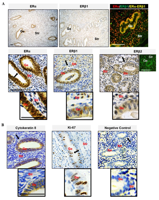
Figure 1.
Localization of ER subtypes in the human endometria. (A) The localization of ERα (red) and ERβ (green). (B) Cellular marker proteins of cytokeratin 8 and Ki 67 in human endometria during the estrogen-dominant proliferative phase was assessed using immunohistology. Sections exposed to human endometrial tissues (the proliferative phase) were used as negative controls. Brown spots were observed using 3,3'-diaminobenzidine as the chromogen. Black arrows indicate areas shown at higher magnification; scale bar, 100 µm. Ge, glandular epithelial cells; Str, stromal cells; ER, estrogen receptor.

