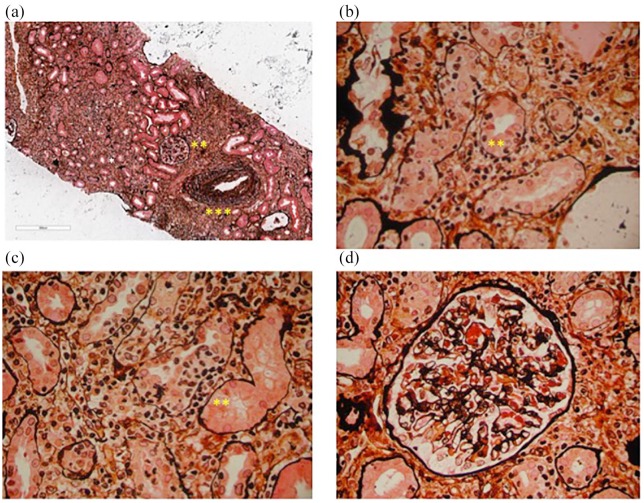
Figure 1.
The hallmark of acute interstitial nephritis induced by an anti-PD-1 monoclonal antibody. (a) A representative renal biopsy from a patient treated with anti-PD-1 who developed severe tubulitis (*). The diffuse infiltrate mostly consists of lymphocytes and plasma cells. The glomerular morphology (**) is almost normal but a mild to moderate intimal fibrosis is seen in an interlobular artery. (b) and (c) Diffuse severe tubular inflammation (*) without findings associated with atrophy, including lymphocyte infiltration and interstitial edema (**). (d) Mild-moderate capillary congestion in a glomerulus surrounded by an interstitial infiltrate but without any other significant abnormalities. Silver methenamine staining of renal tissues visualized by optical microscopy (50× and 400× magnification).

