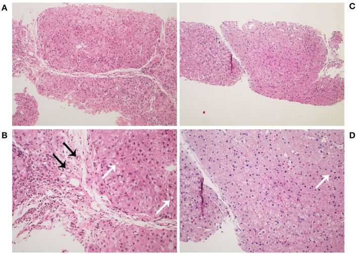
Figure 2.
Histopathology images of liver stained by hematoxylin-eosin. Liver histology in the patient at age 2 years (A,B). It showed a typical picture of cirrhosis with regenerative nodules of hepatocytes separated by fibrous septa (A, ×100). It also shows hydropic degeneration, microvesicular fatty droplets (white arrows), bile duct proliferation (black arrows), and inflammatory cells infiltration (B, ×200). Liver histology in the patient at age 4 years (C,D). It showed lower hydropic degeneration rate and fewer inflammatory cells infiltration.

