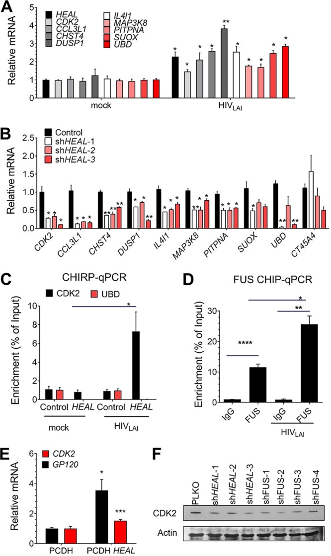
FIG 5.

HEAL is required to maintain expression of CDK2 to support HIV-1 replication. (A) RT-qPCR analysis of HEAL-regulated mRNAs in uninfected or HIV-1-infected MT4 cells 2 days postinfection. Signals were normalized to GAPDH mRNA levels. n = 3, mean ± SD; *, P < 0.05; **, P < 0.01. (B) RT-qPCR analysis of HEAL-regulated mRNAs in MT4 cells expressing control vector or three HEAL shRNAs and then infected with HIV-1. mRNAs were quantified 2 days later. Signals were normalized to GAPDH mRNA levels. n = 3, mean ± SD; *, P < 0.05; **, P < 0.01. (C) qPCR of HEAL-associated DNA in ChIRP assays of uninfected or HIV-1-infected MT4 cells expressing empty vector (control) or overexpressing HEAL. n = 3, mean ± SD; *, P < 0.05. (D) FUS protein interaction with the CDK2 promoter is enhanced by HIV-1 infection. ChIP assays of H9 T cells 7 days after infection with HIV-1. RT-qPCR analysis of the CDK2 promoter region was performed on control IgG or anti-FUS immunoprecipitates. Signals were normalized to GAPDH mRNA levels. n = 3, mean ± SD; *, P < 0.05; **, P < 0.01; ****, P < 0.0001. (E) RT-qPCR analysis of GP120 and CDK2 mRNA in HIV-1-infected MT4 cells expressing empty vector (pCDH) or overexpressing HEAL. Signals were normalized to GAPDH mRNA levels. n = 3, mean ± SD; *, P < 0.05; ***, P < 0.001. (F) Knockdown of HEAL or FUS reduces cellular CDK2 protein levels. MT4 cells were transduced with lentiviruses carrying empty vector or the indicated shRNAs and infected with HIV-1 2 days later. Cell lysates were prepared 2 days after infection and analyzed by Western blotting with anti-CDK2 or anti-β-actin antibodies.
