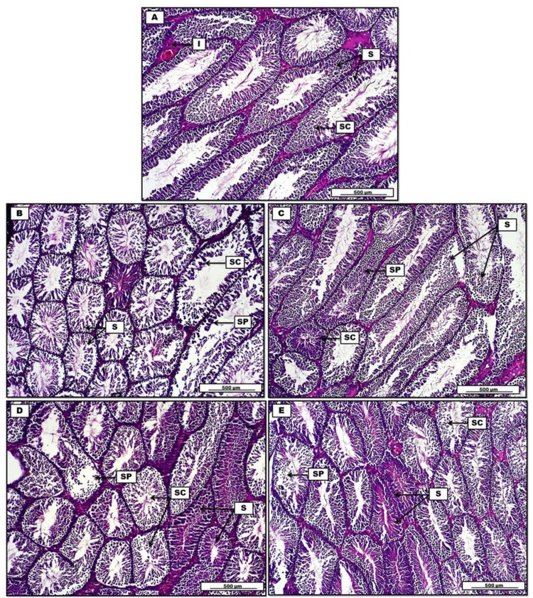
Figure 8.
Photomicrograph of testis from groups; (A) control group, display consistent numbers of seminiferous tubules (S), separated by interstitial connective tissue (I) lined by uniformly arranged spermatogenic cells (SC). (B) Doxorubicin group show a significant reduction in spermatogenesis (SC), decrease in seminiferous tubules spermatogenic cells (S), together with severe degenerative changes in germinative sperm cell layers (SP). (C) Doxorubicin, quercetin and sitagliptin group reveal significant germinal regeneration in the spermatogenic cells (SC). Repopulation in the germinative cells of spermatogenesis (SP). Some tubules show degenerative changes in lining germinal epithelium (S). (D) Doxorubicin and sitagliptin group show regenerative changes in some seminiferous tubules (S), spermatogenic cells (SC) show typical degenerative-atrophy changes, and germinative spermatogenesis epithelium debris within the testicular tubules (SP). (E) Doxorubicin and quercetin group display significant regeneration in seminiferous tubular germinal epithelium (S), marked cellular debris within the tubular lumen (SP), and degenerative changes in spermatogenic cells (SC). H&E. Scale bars: 500 µm.

