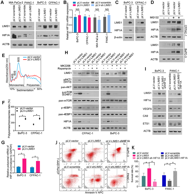
Figure 3. LIMS1 facilitates the response to hypoxic stress by upregulating HIF1A translation.
A-B, the indicated PDAC cells with LIMS1 upregulation or downregulation were subjected to Western blotting (A) and RT-PCR (B) to detect the expression level of HIF1A. C, the indicated PDAC cells were incubated in hypoxic conditions (1% O2) for 48 hours and then subjected to Western blotting to detect the level of HIF1A. D, the indicated PDAC cells were incubated with or without 10 nM MG132 for 24 hours and then subjected to Western blotting to detect the level of HIF1A. E-G, polysome profiles from the indicated cells. Absorbance at 254 nm is shown as a function of sedimentation (E). The area under the curve for polysomes and the 80S peak were calculated, and the ratio is shown (F). The fractions of polysomes were mixed together, and the RNA of the mixture was isolated and subjected to real-time PCR assays to determine the polysomal mRNA level of HIF1A (G). H, the indicated PDAC cells were incubated with or without 10 nM MK2206/0.1 nM rapamycin and then subjected to Western blotting to detect the expression level of p-AKT1, p-mTOR (S2481), p-4EBP1 and HIF1A. The incubation times are as follows: MK2206, 24 hours; rapamycin, 30 minutes for detection of p-4EBP1 and 24 hours for detection of HIF1A. I, the indicated cells were subjected to Western blotting to detect the expression levels of three markers of HIF1 signalling: VEGFA, CA9 and ETS1. J-K, the indicated tumor cells were incubated in hypoxic conditions (oxygen concentration, 0.5%) for 48 hours and then subjected to flow cytometry analysis to detect cell viability. Unpaired t tests were used in B; data are shown as the mean value ± SD; *P < 0.05.

