
Figure 1.
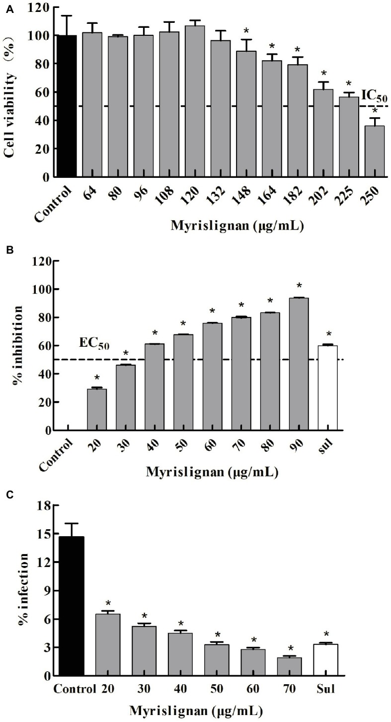
Myrislignan cytotoxicity in Vero cells (A). Vero cells were cultured with myrislignan (64, 80, 96, 108, 120, 132, 148, 164, 182, 202, 225, or 250 μg/ml) or without (control) for 24 h, and the percentages of viable cells were quantified via a CCK-8 assay. All data are presented as the mean ± standard deviation (SD), and the experiments were performed in triplicate. *p < 0.01 compared with the control. Assessment of the anti-proliferation effect of myrislignan on T. gondii-infected Vero cells by qPCR (B). Vero cells were infected with 2.5 × 10⁴ T. gondii for 6 h and then treated with myrislignan (20, 30, 40, 50, 60, 70, 80, or 90 μg/ml), sulfadiazine (0.4 μg/ml) or no drug (parasite control). After 24 h of treatment, total T. gondii DNA in each group was extracted, and the 529-bp repeat element of the T. gondii genome was measured by PCR. The percent inhibition of tachyzoite proliferation in each group was compared with that in the parasite control group. The data are presented as the mean ± SD of three independent experiments. *p < 0.01 compared with the parasite control. Effects of myrislignan on T. gondii invasion (C). Vero cells were cultured in DMEM with myrislignan (20, 30, 40, 50, 60, or 70 μg/ml), sulfadiazine (0.4 μg/ml), or no drug (parasite control) and infected with 2.5 × 10⁶ T. gondii tachyzoites. After 2 h of infection, the cells were subjected to Giemsa staining, and the percentage of infected cells in each group was determined by light microscopy. At least 15 random fields per well were counted. Triplicate independent experiments were performed, and the data are presented as the mean ± SD. *p < 0.01 compared with the parasite control.