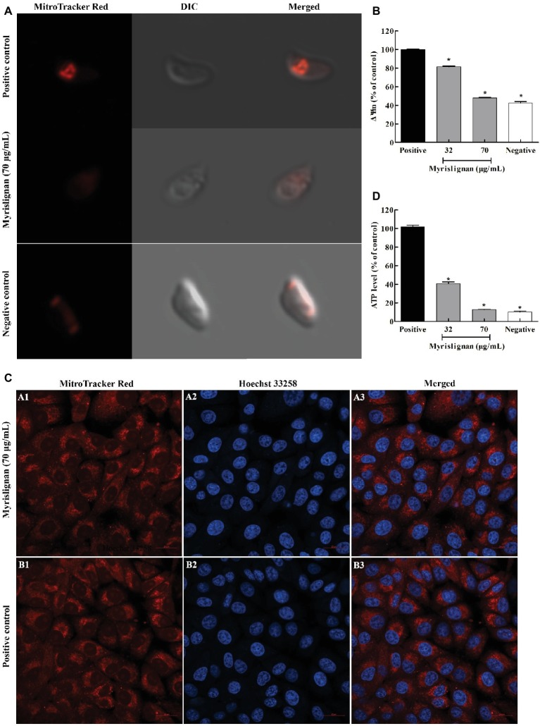
Figure 5.
Myrislignan decreased the ΔΨm and ATP levels of T. gondii tachyzoites. T. gondii tachyzoites (1 × 10⁵ per group) were incubated with myrislignan (32 or 70 μg/ml) or with no drug (positive or negative control) in DMEM for 8 h at 37°C. The negative control samples were further incubated with 10 μM CCCP for 20 min. All the samples were then stained with MitoTracker Red CMXRos for 20 min at 37°C in the dark, rinsed twice with PBS, centrifuged and suspended in 500 μl of PBS. The fluorescence intensity of each group was then observed by laser confocal microscopy (A). The fluorescence intensities of the positive or negative control group and 32 μg/ml and 70 μg/ml myrislignan treatment groups were assessed using a multilabel reader (B). Evaluation of the effect of myrislignan on the mitochondria of Vero cells (C). Vero cells were placed in cell culture dishes and incubated with myrislignan (70 μg/ml) in DMEM or without drug (positive control) for 8 h at 37°C, stained with the MitoTracker Red CMXRos probe (250 nM) for 20 min at 37°C, rinsed twice with PBS, fixed with 4% polyformaldehyde for 15 min, washed twice with PBS to remove the polyformaldehyde, stained with Hoechst 33342 and then observed by laser confocal microscopy. Myrislignan induced a decrease in the ATP concentration (D). T. gondii tachyzoites (1 × 10⁶ per group) were incubated with myrislignan (32 or 70 μg/ml) or without any drug (parasite positive or negative control) in DMEM for 8 h at 37°C. The negative control samples were then incubated with CCCP. The ATP levels of T. gondii tachyzoites were determined using a multilabel reader. The ATP levels are expressed as a percentage of the positive control. Data are presented as the mean value ± SD from three replicate experiments. *p < 0.01 compared with the positive parasite control.

