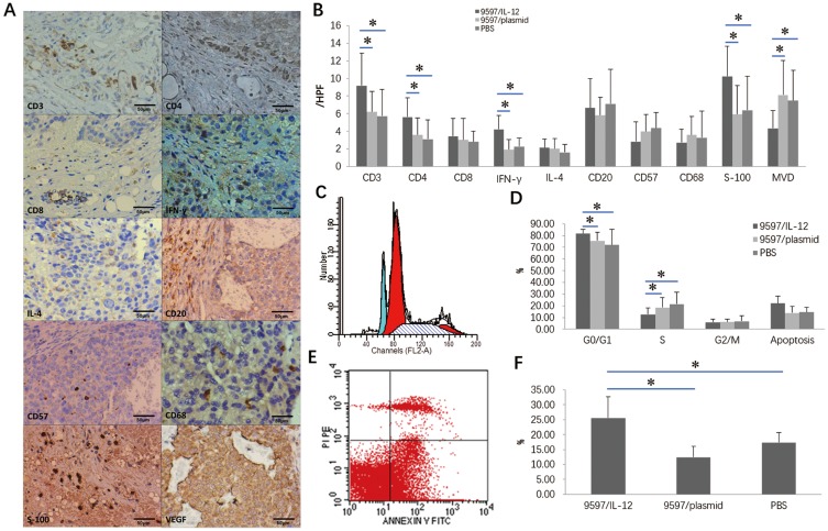
Figure 5.
Intratumoral 9597/IL-12 cell injection altered tumor microenvironments, cell cycle and apoptosis. (A) IHC detects expression of CD3+T cells, CD4+T helper cells, IFN-γ Th1 cells+ and S-100 protein positive DCs and MVD in tumor microenvironments (scale bar = 50 μm); (B) higher counts of CD3+T cells, CD4+T helper cells, IFN-γ Th1 cells+ and S-100 protein positive dentric cells (DCs) in the 9597/IL-12 group than in the 9597/plasmid and PBS groups, and the MVD was significantly lower in the 9597/IL-12 group than in the 9597/plasmid and PBS groups; (C) Flow cytometry detects cell cycle in tumor microenvironments; (D) A significantly higher proportion of HCC cells at the G0/G1 phase and a significantly lower proportion of HCC cells at the S phase are seen in the 9597/IL-12 group than in the PBS group, and no significant difference is detected in the proportion of HCC cells at the G2/M phase among the three groups; (E) Flow cytometry detects cell apoptosis in tumor microenvironments; (F) A greater apoptotic rate of HCC cells is determined in the 9597/IL-12 group than in the 9597/plasmid and PBS groups. *P < 0.05.

