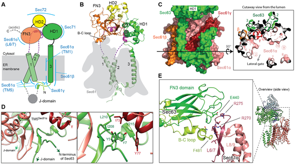
Figure 2. Structure of Sec63 and interactions with the channel.
(A) A schematic of Sec63 domains. Regions interacting with other parts of the complex are indicated by blue lines. Unmodeled regions are shown in dashed lines. (B) Structure of Sec63 (front view). The position of Sec61 is shown by a gray shade. (C) Interactions between TMs of Sec63 and Sec61. Left, a view from the back; right, a cutaway view from the ER lumen. Black arrowed line, the cross-sectional plane. Note that TMs 2, 9, and 10 of Sec61α are located above the cross-sectional plane. (D) Interactions between Sec63 and Sec61 in the luminal side. Left, a β-sheet formed between Sec61α (TM5 indicated by a dashed line) and the segment between Sec63 TM3 and the J-domain. Right, a magnified view with side chains in sticks. (E) Interactions between the FN3 domain and the cytosolic loop L6/7 of Sec61α (also see Fig. 1B).

