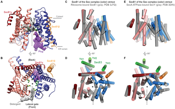
Figure 3. A fully opened Sec61 channel in the Sec complex.
(A and B) Structure of the Sec61 channel. The N- and C- terminal halves of Sec61α are in blue and salmon, respectively. Gray density feature is presumed detergent molecules. Pore-lining residues are shown as green balls and sticks. Density feature for the plug is in purple. ‘2’ and ‘7’ indicate TM2 and TM7 respectively. (C–F) Comparison of Sec61 of the Sec complex (colored) with Sec61 of the cotranslational ribosome-Sec61 complex (gray; C and D) or SecY of a bacterial posttranslational SecA-SecY channel complex (gray; E and F). The structures are aligned with respect to the C-terminal half of Sec61α (C–F). Shown are the front (A, C, and E) and cytosolic (B, D, and F) views. Numbers indicate corresponding TMs. Dashed line, lateral gate. Asterisk, translocation pore. For simplicity, L6/7 and L8/9 of Sec61α were not shown. In D and F, TMs of Sec63 are also shown (green). Also see fig. S6 for comparisons to archaeal SecY and substrate-engaged channels.

