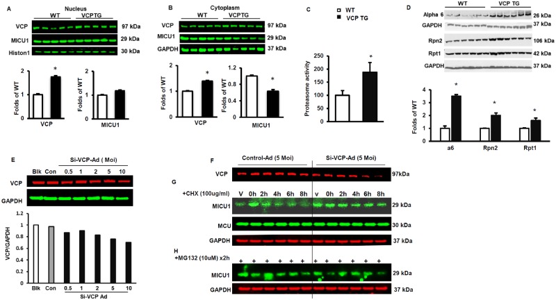
Figure 6.
Valosin-containing protein (VCP) increases MICU1 degradation via proteasome. A and B, Subcellular distribution of MICU1 in the mouse heart tissue. Western blot showing protein expression of VCP and MICU1 in nuclear fractions (A) and cytoplasmic fractions (B) *p < .05 versus wild type (WT). N = 5/group. C, Overexpression of VCP increases proteasome activity in heart tissues of VCP transgenic (TG) mice versus WT mice. N = 4/group with triplication in each samples *p < .05 versus WT. D, Western blot showing protein expression of key components of the Proteasome: a6, Rpn2, and Rpt 1. N = 6/group. *p < .05 versus WT. E, Western blotting showing a dose response of the reduction of VCP in H9ce cell upon the knock down of VCP by a Si-VCP adenovirus versus control. F, VCP protein levels in H9C2 cells upon the addition of 5 MOI Si-VCP Ad. G, A cycloheximide (CHX)-chase assay showing half-life of MICU1 and MCU in H9C2 cells upon 5 MOI Si-VCP Ad versus controls. H, Representative Western blots showing MICU1 levels with the treatment of proteasome inhibitor (MG132) in CHX-treated cells for 2 h. Glyceraldehyde 3-phosphate dehydrogenase (GAPDH) was used as loading control of the total proteins from the cells.

