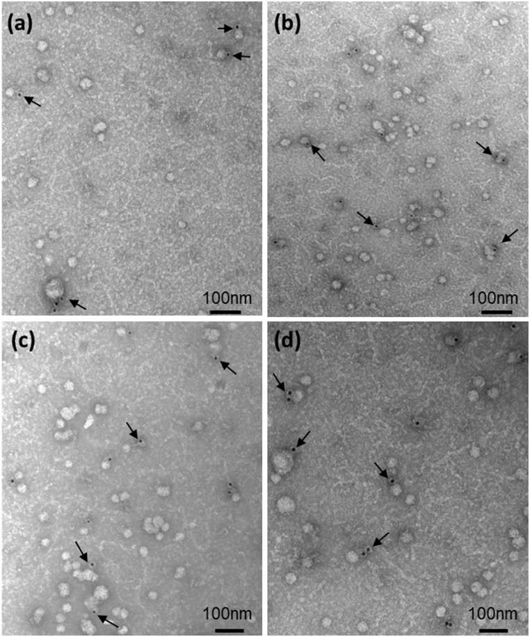
Figure 2.
Transmission electron microscopy images of EVs isolated from the serum of (a and b) two controls and (c and d) two women with GDM. The presence of EVs of placental origin was confirmed by immunostaining with mouse monoclonal antihuman-PLAP IgG followed by 12 nm Colloidal Gold AffiniPure goat anti-mouse IgG (Cat No. 115-205-166; Jackson ImmunoResearch Laboratories Inc). Samples were stained with uranyl acetate. Arrows point to gold particles linked to the antibody anti-PLAP. Scale bar = 100 nm.

